
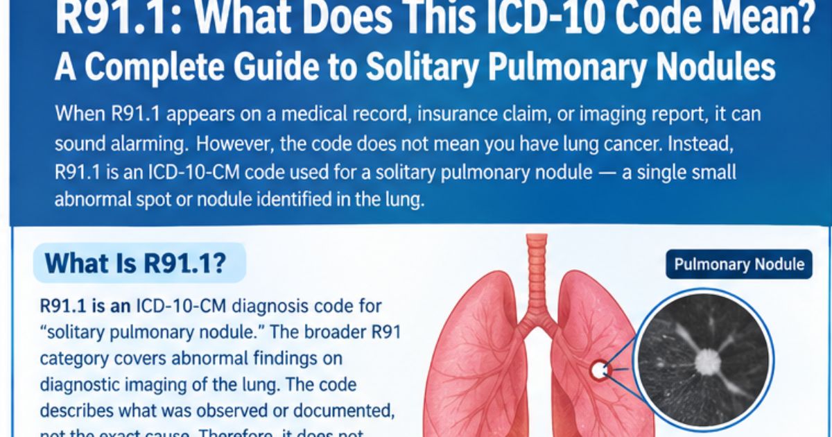
When R91.1 appears on a medical record, insurance claim, or imaging-related document, it can sound alarming. However, the code itself does not mean that you have lung cancer. Instead, R91.1 is an ICD-10-CM code used for a solitary pulmonary nodule, meaning a single small abnormal spot or nodule identified in the lung. Therefore, the code describes an imaging finding rather than automatically identifying its cause. Moreover, lung nodules can develop for many reasons, including previous infections, inflammation, scar tissue, or other conditions. Consequently, doctors usually consider the nodule’s size, appearance, location, growth, and the patient’s individual risk factors before deciding what should happen next.
What Is R91.1?
R91.1 is an ICD-10-CM diagnosis code for “solitary pulmonary nodule.” The broader R91 category covers abnormal findings on diagnostic imaging of the lung. In addition, the R91.1 description includes terms such as a coin lesion of the lung and a solitary pulmonary nodule located in a subsegmental branch of the bronchial tree. Because ICD-10-CM codes classify medical findings and diagnoses in a standardized way, the code helps healthcare professionals communicate information consistently. Importantly, R91.1 does not by itself establish why the nodule exists. Instead, it records what was observed or documented. Therefore, patients should read the code together with the radiology report and their doctor’s explanation rather than interpreting the code as a final diagnosis of cancer or another specific disease.
R91.1 and the Meaning of a Pulmonary Nodule
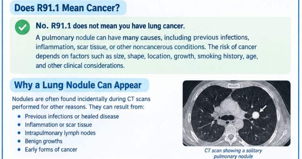
A pulmonary nodule is generally a small, rounded or irregular area seen within the lung on imaging, particularly CT scans. Often, these nodules are discovered unexpectedly when a person receives chest imaging for another reason. For example, a scan may be performed because of coughing, chest discomfort, an infection, or an unrelated medical evaluation. Subsequently, the radiologist may identify a small spot that requires further attention. However, a nodule is not automatically dangerous. Many nodules are benign, especially when they have reassuring features. Nevertheless, some nodules require monitoring because certain characteristics can increase concern. Therefore, doctors evaluate the complete clinical picture rather than relying on the R91.1 code alone.
Does R91.1 Mean Cancer?
No. R91.1 does not mean that a person has lung cancer. Instead, it identifies a solitary pulmonary nodule that has been detected or documented. That distinction is extremely important because imaging can reveal an abnormal spot without immediately determining whether it is benign or malignant. Furthermore, the probability of cancer depends on several factors, including nodule size, shape, location, growth pattern, smoking history, age, and other clinical considerations. The Fleischner Society guidelines specifically emphasize risk-based management of incidental pulmonary nodules. Therefore, someone who sees R91.1 on a report should avoid assuming the worst. Instead, the next step should be a discussion with the healthcare professional who can interpret the imaging in its proper clinical context.
Why a Lung Nodule Can Appear on a CT Scan
Lung nodules can have numerous causes, and many are unrelated to cancer. For instance, a previous infection can leave a small area of scar tissue or inflammation that remains visible on imaging. Similarly, some nodules can represent healed granulomatous disease or intrapulmonary lymph nodes. Additionally, inflammatory conditions and other noncancerous processes can create pulmonary nodules. Consequently, the discovery of a nodule should be viewed as a finding that requires interpretation rather than a definitive disease diagnosis. Moreover, modern CT technology can detect very small abnormalities that might previously have gone unnoticed. As a result, incidental pulmonary nodules are encountered relatively often, and careful assessment helps doctors avoid unnecessary procedures while still identifying potentially important findings.
How Doctors Evaluate an R91.1 Finding
When R91.1 is documented, doctors generally begin by reviewing the imaging report and, when appropriate, the actual CT images. They may examine the nodule’s size, density, shape, margins, location, and appearance. Furthermore, they may compare the current scan with older images to determine whether the nodule has changed over time. Stability can provide important information, although the meaning of stability depends on the type and characteristics of the nodule. In addition, clinicians consider personal risk factors and medical history. The Fleischner Society recommends that incidental pulmonary nodule management incorporate factors such as nodule characteristics and the patient’s estimated risk. Therefore, two people with the same R91.1 code may receive different follow-up recommendations.
Why Nodule Size Matters
Size is one of the important factors doctors consider when evaluating a solitary pulmonary nodule. According to the 2017 Fleischner Society recommendations, management of a single solid nodule differs according to whether it measures less than 6 mm, 6–8 mm, or more than 8 mm. For example, routine follow-up may not be necessary for some very small nodules in people considered low risk. Conversely, a larger nodule may require additional imaging or further evaluation. However, size should never be considered in isolation. A smaller nodule with suspicious characteristics may deserve attention, while a larger nodule with reassuring features may be managed differently. Therefore, the radiologist’s description and the patient’s clinical risk profile remain essential when interpreting an R91.1 finding.
R91.1 and Follow-Up CT Scans
Follow-up imaging is sometimes recommended after a solitary pulmonary nodule is discovered. However, the timing depends on the nodule’s size, type, appearance, and the patient’s risk factors. For example, the Fleischner Society recommends that some solitary solid nodules measuring 6–8 mm receive CT follow-up, while management of nodules larger than 8 mm can include several possible approaches. Furthermore, subsolid nodules can require a different surveillance strategy because they may behave differently from solid nodules. Consequently, there is no universal R91.1 follow-up schedule that applies to every person. Instead, the doctor should interpret the imaging characteristics and determine an appropriate plan. Therefore, patients should follow the specific schedule provided by their healthcare team rather than relying on a generic timetable.
What Happens If the Nodule Does Not Change?
If a pulmonary nodule remains stable on appropriate follow-up imaging, that information can be reassuring. Nevertheless, the significance of stability depends on the nodule’s characteristics and type. For certain solid nodules, the Fleischner Society guidelines explain that stability over an appropriate period can support a benign interpretation. However, subsolid nodules may require longer surveillance because some can change slowly. Therefore, doctors do not always apply the same follow-up period to every nodule. Additionally, accurate comparison requires suitable imaging and consistent measurement. Consequently, patients should not assume that one unchanged scan automatically ends follow-up. Instead, the treating clinician should determine whether additional imaging is appropriate based on the complete clinical and radiological picture.
What Is a Solitary Pulmonary Nodule?
The word solitary in R91.1 is significant because it indicates a single pulmonary nodule rather than multiple nodules. A solitary pulmonary nodule can be solid or subsolid, depending on how it appears on CT imaging. Furthermore, its appearance can provide clues about possible causes. For example, a smooth, well-defined nodule may have different implications from a lesion with irregular or spiculated margins. Location can also influence risk assessment. Nevertheless, no single imaging characteristic can reliably diagnose cancer on its own. Therefore, radiologists combine multiple observations when preparing an imaging report. Ultimately, the R91.1 code provides a standardized label for the finding, while the detailed radiology report provides the information needed for meaningful clinical interpretation and follow-up planning.
Solid and Subsolid Nodules
Pulmonary nodules can have different appearances on CT scans. A solid nodule appears relatively dense throughout its visible area, whereas a subsolid nodule may include ground-glass or part-solid components. These distinctions matter because follow-up recommendations can differ substantially. According to the Fleischner Society, solitary solid and subsolid nodules have separate management considerations based on their size and appearance. For example, persistent subsolid nodules may require longer surveillance than certain small solid nodules. Therefore, a patient should look beyond the R91.1 code and review the wording of the radiology report. Moreover, terms such as “solid,” “ground-glass,” “part-solid,” “spiculated,” and “stable” can provide important context for the treating physician.
What Causes a Solitary Pulmonary Nodule?
A solitary pulmonary nodule can result from several conditions. Previous infections are among the possible benign causes, particularly when inflammation or healed disease leaves a visible area in the lung. In addition, benign growths, scars, inflammatory changes, and intrapulmonary lymph nodes can appear as nodules. At the same time, some pulmonary nodules can represent an early form of cancer. Therefore, doctors do not dismiss every nodule, but they also do not assume every nodule is malignant. Instead, they estimate the likelihood of different causes using imaging findings, medical history, risk factors, and changes over time. Consequently, an R91.1 finding should prompt appropriate medical evaluation rather than immediate fear or assumptions.
Risk Factors Doctors May Consider
Doctors may consider several factors when estimating the likelihood that a pulmonary nodule could represent cancer. Smoking history is particularly important because tobacco exposure increases lung cancer risk. Additionally, age, family history, nodule location, morphology, and certain underlying lung conditions can influence clinical assessment. The Fleischner recommendations identify factors such as tobacco smoking, family history of lung cancer, suspicious nodule shape, upper-lobe location, rapid growth, emphysema, and fibrosis among considerations related to higher risk. However, these factors do not independently diagnose cancer. Instead, they help clinicians decide whether closer monitoring or additional testing may be appropriate. Therefore, patients should provide their healthcare professional with a complete and accurate medical history.
Can an R91.1 Nodule Go Away?
Yes, some pulmonary nodules or lung opacities can disappear or decrease in size, particularly when they result from temporary inflammation or infection. This possibility is one reason why doctors sometimes recommend follow-up imaging rather than immediately performing an invasive procedure. For instance, the Fleischner Society notes that some subsolid nodules can represent transient infectious or inflammatory processes and may resolve during short-term follow-up. Nevertheless, disappearance is not something a patient should assume will happen. Similarly, persistence does not automatically mean cancer. Instead, changes over time provide one piece of evidence within a broader assessment. Consequently, follow-up should occur according to the medical plan, particularly when the original imaging report recommends repeat CT imaging or specialist review.
When Can Additional Testing Be Needed?
Additional testing may be considered when a nodule has characteristics that make further evaluation appropriate. For example, the Fleischner Society recommends considering short-term CT, PET/CT, tissue sampling, or combinations of these approaches for certain solitary solid nodules larger than 8 mm. However, that recommendation does not mean every nodule above that size requires a biopsy or PET scan. Instead, doctors consider morphology, size, medical history, risk factors, and overall health. Furthermore, invasive testing has its own risks, so clinicians generally balance potential benefits against possible harms. Therefore, a patient with R91.1 should not request or assume a particular test solely because of the code. The imaging findings and clinical assessment should guide the decision.
Why PET/CT or Biopsy May Be Considered
PET/CT and tissue sampling can sometimes provide additional information when imaging alone does not adequately characterize a pulmonary nodule. A PET/CT can evaluate metabolic activity, while biopsy can allow tissue to be examined directly. However, neither test is automatically appropriate for every R91.1 finding. The Fleischner recommendations specifically present PET/CT and tissue sampling as possible options for selected larger or concerning nodules rather than universal requirements. Therefore, doctors first assess whether additional testing is likely to change management. Moreover, factors such as nodule location, patient health, procedural risks, and imaging appearance can influence the choice. Consequently, patients should discuss the purpose, benefits, limitations, and risks of any recommended test before proceeding.
R91.1 in Medical Coding and Health Records
From a coding perspective, R91.1 belongs to the ICD-10-CM classification system. The CDC explains that ICD-10-CM is used to code and classify medical diagnoses and related health information in the United States. Therefore, the code can appear in electronic health records, billing documentation, claims, or clinical records. Importantly, coding terminology and clinical interpretation are not identical. A code may describe an observed finding while the underlying cause remains under evaluation. For that reason, seeing R91.1 in an online health portal does not necessarily mean that a definitive disease has been diagnosed. Instead, patients should review the accompanying documentation, including the radiology impression and any assessment or plan provided by their healthcare professional.
Is R91.1 a Billable Code?
For the 2026 ICD-10-CM classification, R91.1 is listed as a billable diagnosis code for a solitary pulmonary nodule. The code is classified under abnormal findings on diagnostic imaging of the lung. Furthermore, coding references identify “coin lesion lung” as an inclusion term associated with R91.1. However, being billable does not make the code a diagnosis of cancer or another specific disease. Instead, it provides a standardized way to document the pulmonary finding. Therefore, patients should distinguish between the administrative purpose of the code and the clinical meaning of the imaging finding. Ultimately, the radiologist’s report and physician’s assessment provide much more context than the code alone.
Questions to Ask Your Doctor About R91.1
After receiving an R91.1 finding, patients may feel uncertain about what to ask next. Fortunately, a short list of focused questions can make the discussion more productive. For example, ask how large the nodule is, whether it is solid or subsolid, where it is located, and whether it has changed compared with previous scans. Furthermore, ask whether your personal risk factors affect the recommended follow-up plan. You can also ask why another CT scan, PET/CT, or biopsy is or is not being considered. Because management depends on individual circumstances, these questions can help you understand the reasoning behind your doctor’s recommendation. Consequently, informed communication can reduce unnecessary anxiety while ensuring that important follow-up is not overlooked.
When Should You Seek Prompt Medical Advice?
R91.1 itself does not describe an emergency. Nevertheless, a person should follow medical advice if a lung nodule has been identified, especially when follow-up imaging has been recommended. Additionally, new or worsening respiratory symptoms should be discussed with a healthcare professional rather than automatically attributed to the nodule. Symptoms such as significant breathing difficulty, coughing up blood, severe chest pain, or rapidly worsening illness can have many causes and may require prompt assessment. Importantly, symptoms cannot determine whether an R91.1 finding is benign or malignant. Therefore, do not use online information to replace professional evaluation. Instead, provide your clinician with the complete imaging report, previous scans when available, medication history, smoking history, and relevant medical information.
R91.1: Key Facts to Remember
The most important point is simple: R91.1 means solitary pulmonary nodule, not lung cancer. The code documents an imaging finding that requires appropriate interpretation. Furthermore, the significance of that finding depends on characteristics such as size, density, shape, location, growth, and the patient’s individual risk profile. Some nodules are caused by previous infections or other benign processes, while others may require closer investigation. Therefore, follow-up recommendations can differ considerably between patients. Moreover, established guidelines emphasize individualized risk assessment rather than a one-size-fits-all approach. If you have received this code, review the complete radiology report and discuss the recommended next step with your healthcare professional instead of assuming that the code represents a final diagnosis.
Frequently Asked Questions About R91.1
What does R91.1 mean?
R91.1 means “solitary pulmonary nodule” in ICD-10-CM. It describes a single nodule or abnormal spot identified in the lung, usually through imaging. Therefore, it is an imaging finding rather than a statement that the patient has cancer.
Is R91.1 the same as lung cancer?
No. R91.1 is not a lung cancer diagnosis. Instead, it identifies a solitary pulmonary nodule whose cause may still need evaluation. Some nodules are benign, while others may require additional investigation. Consequently, the radiology report and clinical assessment are essential.
What causes an R91.1 pulmonary nodule?
Several causes are possible. Previous infections, inflammation, scar tissue, intrapulmonary lymph nodes, benign growths, and cancer can all be considered depending on the imaging appearance and clinical circumstances. Therefore, additional context is needed before identifying the cause.
Does every R91.1 nodule require a biopsy?
No. A biopsy is not automatically required. Management depends on factors such as size, appearance, growth, location, and patient risk. For some nodules, doctors may recommend surveillance CT instead. For selected larger or concerning nodules, PET/CT or tissue sampling may be considered.
How is an R91.1 nodule monitored?
Monitoring may involve repeat CT imaging at an interval selected according to the nodule’s characteristics and the patient’s risk. The Fleischner Society provides recommendations for different nodule sizes and types. However, those recommendations do not apply to every clinical situation, so individualized medical advice remains important.
Can an R91.1 nodule disappear?
Yes, some nodules or similar lung findings can resolve, particularly when they are related to temporary infection or inflammation. Nevertheless, persistence does not automatically mean cancer. Doctors use follow-up imaging and other clinical information to determine what the finding means.
What should I do if R91.1 appears on my report?
First, do not panic or assume that you have cancer. Instead, obtain the complete radiology report and ask your healthcare professional about the nodule’s size, type, location, appearance, and recommended follow-up. Additionally, mention previous scans and relevant risk factors so the doctor can make an informed assessment.
Conclusion: Understand R91.1 Before You Assume the Worst
R91.1 is an ICD-10-CM code for a solitary pulmonary nodule. Although seeing a lung-related code can understandably cause concern, the code itself does not establish cancer. Instead, it records an imaging finding that needs to be interpreted in context. Furthermore, many pulmonary nodules have benign explanations, while certain features may require closer monitoring or additional testing. Therefore, the smartest next step is not to guess from the code but to review the complete imaging report with a qualified healthcare professional. Ask about the nodule’s size, appearance, growth, risk factors, and recommended follow-up. If you or someone you know has received an R91.1 finding, use this information as a starting point and arrange an appropriate discussion with your healthcare provider.









2 Comments